Podcast

 Rebecca Previs

Biomarkers have moved from the margins of oncology to the center of clinical decision-making. They now help establish diagnosis and prognosis, identify patients most likely to benefit from targeted therapy or immunotherapy, monitor response, detect molecular residual disease and emerging resistance, and anticipate selected toxicities. At the same time, the sources of biomarker information are expanding beyond tumor tissue. Circulating tumor DNA (ctDNA), quantitative imaging, digital pathology, wearable sensors, and remotely collected patient data are creating the possibility of a more continuous view of both the cancer and the person living with it. This narrative review presents a practical framework for understanding predictive, monitoring, safety, and digital biomarkers; highlights their application in precision therapeutics, immuno-oncology, antibody-drug conjugates, precision radiation oncology, theranostics, and post-treatment surveillance; and examines the growing role of artificial intelligence, radiomics, and spatial biology in integrating molecular, imaging, pathologic, physiologic, and clinical signals. The central challenge is no longer simply whether a signal can be measured. It is whether the measurement is analytically reliable, clinically valid, prospectively useful, equitable, interpretable, and linked to a decision that improves outcomes. The next phase of precision oncology will therefore be defined less by any single assay than by longitudinal, multimodal biomarker strategies that convert complex data into timely, patient-centered action.
AUTHORS:
David J. Segarnick, PhD
Chief Medical Officer, Educational Resource Systems
Adjunct Associate Professor, Rutgers New Jersey Medical School
Department of Pharmacology, Physiology & Neuroscience
David E. Wazer, MD, FACRO, FABS, FACR, FASTRO
Research Scholar Professor
Department of Radiation Oncology
The Alpert Medical School of Brown University
Providence, RI
Ask an oncologist what has changed cancer care most over the past 2 decades and the answer may be a drug, a technology, or an entirely new treatment class. Running through all 3, however, is the same enabling force: the biomarker. Cancer is still defined by its organ of origin and histologic appearance, but those features are now joined by molecular and immune characteristics that can divide one tumor type into biologically and therapeutically distinct diseases. The reverse is also true: the same actionable alteration may appear across several tumor types, opening the door to histology-agnostic treatment. Biomarker testing now informs care across most major malignancies and a growing number of tumor-agnostic indications.1-5
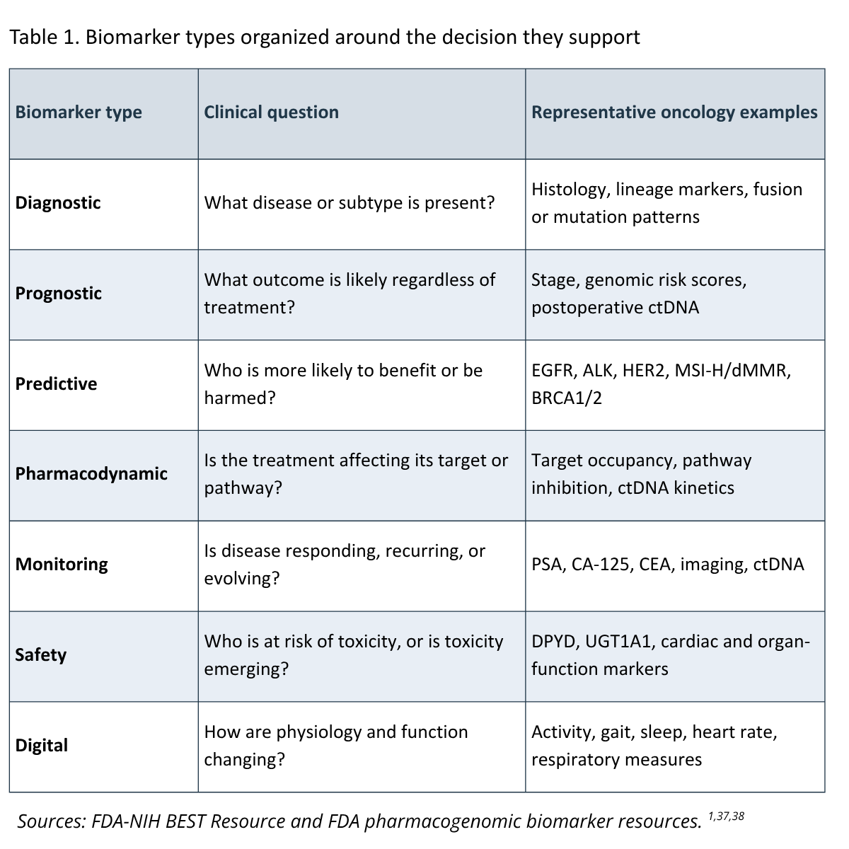
The term biomarker is broad. The FDA NIH Biomarkers, EndpointS, and other Tools (BEST) resource defines it as a measured characteristic that indicates a biologic or pathogenic process or a response to an exposure or intervention.1 For the oncology team, the more useful question is simpler: what decision will this result help us make? A biomarker may help identify the disease, estimate prognosis, select therapy, demonstrate target engagement, monitor response, detect recurrence, or signal toxicity. The same marker can play more than one role, and its meaning can change with tumor type, disease stage, specimen, assay, cutoff, and clinical context.
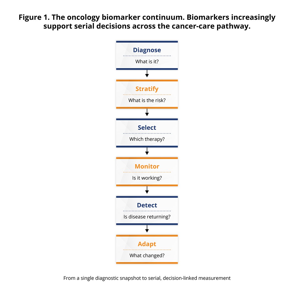
This review organizes the expanding field around 3 practical functions. Predictive biomarkers help match a patient to a treatment. Monitoring biomarkers help determine whether the disease is responding, recurring, or evolving. Digital biomarkers provide objective, digitally acquired measures of physiology, function, or behavior between conventional assessments. Across all 3, oncology is moving from a static model—one biopsy, one test, one decision—toward a longitudinal model in which serial tissue, blood, imaging, functional, and patient-experience data are interpreted together.2
The BEST framework separates biomarkers from clinical outcome assessments and from surrogate endpoints.1 This distinction is crucial. A reduction in ctDNA, improvement in a radiomic feature, or increase in daily step count may be biologically meaningful, but it is not automatically equivalent to longer survival, symptom relief, improved quality of life, or preserved function. A biomarker becomes a surrogate endpoint only when evidence supports using its effect to predict a clinical benefit in a defined context.
A useful way to cut through the complexity is to ask 3 questions in sequence. First, can the assay measure the signal accurately and reproducibly? Second, is that signal reliably associated with the disease, outcome, treatment response, or toxicity of interest? Third—and most importantly—does using the result lead to a better decision or outcome? Regulatory approval of a companion diagnostic answers these questions for a specific drug-test context. It does not make every platform, specimen type, or threshold interchangeable.2,3
This framework also explains why an apparently strong biomarker may fail in practice. Preanalytic variables can alter tissue quality or circulating DNA yield. Assays may use different antibodies, sequencing panels, bioinformatic pipelines, scoring systems, or thresholds. Tumor heterogeneity can make a single biopsy unrepresentative, while therapy can change biomarker status over time. Finally, even a technically accurate result may have limited value if turnaround time is too slow, the result is not available at the treatment decision, or no evidence-based action follows.
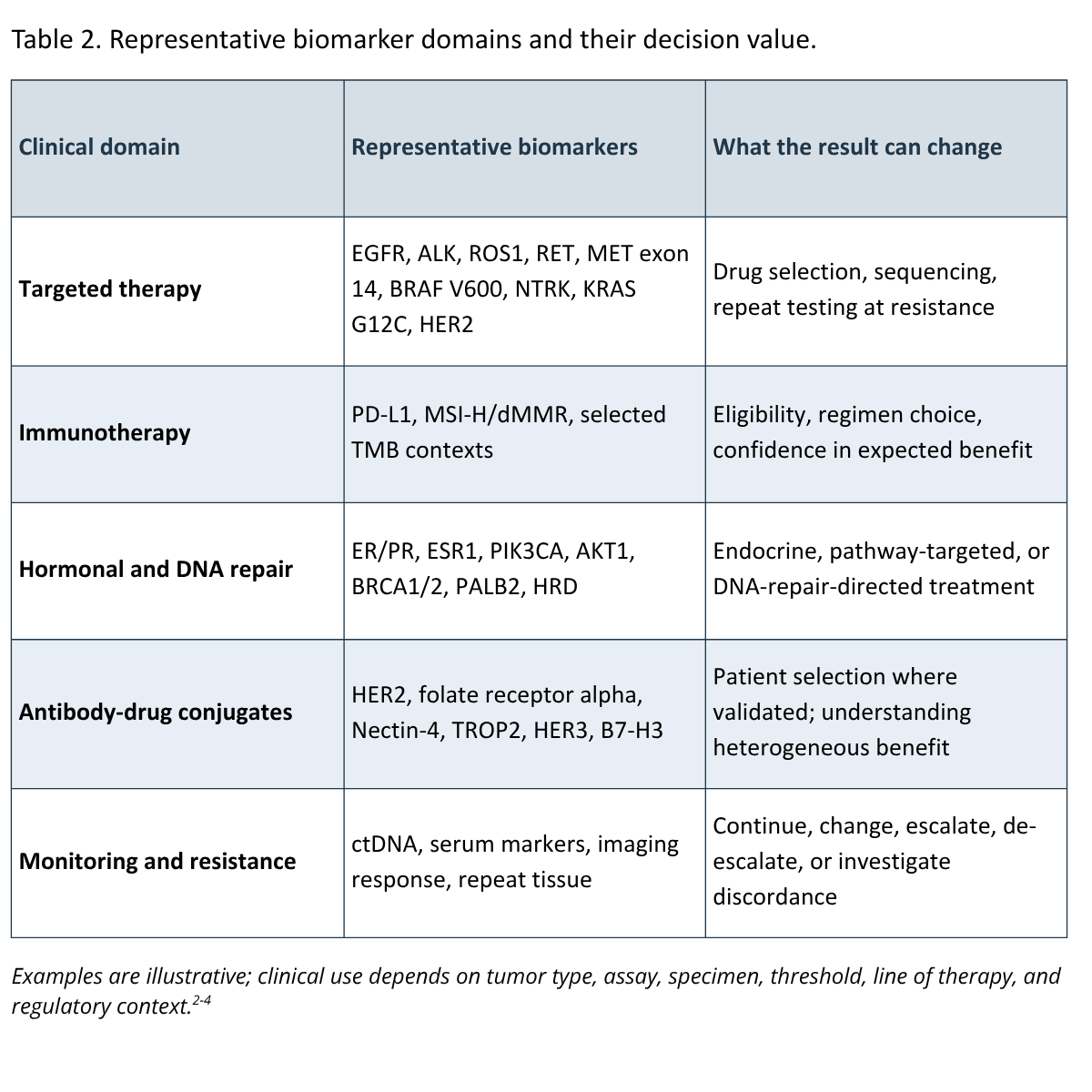
Predictive biomarkers: turning tumor biology into a treatment choicePredictive biomarkers are precision oncology's most visible success story: find the alteration, match the therapy, and change the odds for the patient. Their impact is clearest when a molecular alteration is biologically linked to a drug target. Examples include EGFR mutations, ALK and ROS1 rearrangements, RET fusions, MET exon 14 skipping, BRAF V600 alterations, NTRK fusions, HER2 amplification or mutation, KRAS G12C, IDH1/2 mutations, and selected FGFR alterations. Trials of osimertinib in EGFR-mutated non-small cell lung cancer, alectinib in ALK-positive disease, and larotrectinib in NTRK fusion-positive tumors show how sharply a biomarker can reshape the expected benefit of treatment.6-8
Predictive testing has also expanded beyond single oncogenic drivers. In breast and gynecologic cancers, estrogen and progesterone receptors, HER2, PIK3CA, AKT1, PTEN, ESR1, BRCA1/2, PALB2, and homologous recombination deficiency can influence treatment selection. In immuno-oncology, several biomarkers can identify patients more likely to benefit from immune checkpoint inhibitors: mismatch repair deficiency (dMMR), microsatellite instability-high status (MSI-H), PD-L1 expression, and, in selected settings, high tumor mutational burden (TMB). The activity of pembrolizumab in dMMR/MSI-H tumors helped establish one of the first histology-agnostic treatment paradigms, and randomized data subsequently confirmed the value of first-line checkpoint inhibition in MSI-H metastatic colorectal cancer.9,10
The apparent simplicity can be misleading. Biomarker positivity is not a guarantee of response. A driver may be present in only part of the tumor; a co-mutation may activate a bypass pathway; drug delivery may be inadequate; and resistance may exist at baseline or emerge quickly. The right interpretation is probabilistic: the biomarker changes the likelihood of benefit—sometimes dramatically—but rarely determines the outcome by itself.
Testing strategy matters as well. Single-gene assays may be efficient when one high-prevalence alteration dominates a treatment decision, whereas broad next-generation sequencing can identify multiple actionable changes, resistance alterations, germline implications, and trial options from limited tissue. Tissue remains essential for histology, architecture, protein expression, and many copy-number or fusion assessments. Plasma genotyping can shorten turnaround and sample multiple metastatic sites, but a negative plasma result may reflect low tumor shedding rather than true absence of the alteration. When clinical suspicion remains high, a negative liquid biopsy may need tissue confirmation.2,4
Choosing treatment is only the first decision. The next questions arrive quickly: Is it working? Is the response durable? Is resistance beginning before it is visible on a scan? Traditional serum tumor markers —including prostate-specific antigen, CA-125, carcinoembryonic antigen, CA 19-9, alpha-fetoprotein, beta-human chorionic gonadotropin, lactate dehydrogenase, monoclonal protein, and free light chains—remain valuable in selected malignancies.15 Their limitations are familiar: imperfect sensitivity and specificity, nonmalignant causes of elevation, discordance with radiographic burden, and variable performance across individuals. Their greatest value usually comes from the pattern over time, interpreted in the right disease context, rather than from one isolated result.
Imaging remains the foundation of response assessment in solid tumors. RECIST 1.1 provides a standardized method for measuring changes in target lesions, while immune-specific criteria such as iRECIST were developed to address atypical patterns including apparent progression followed by response.13,14 Yet conventional imaging is an anatomic snapshot. It may not detect early biologic response, can be confounded by inflammation or treatment-related change, and can average away heterogeneous responses across lesions. Pathologic complete response and major pathologic response add important information in neoadjuvant settings, but they are available only when sufficient tissue is obtained.
ctDNA offers a noninvasive window into tumor burden and evolution. It can be assessed through tumor-informed assays that track variants identified in an individual's tumor, or tumor-naive assays that detect predefined genomic and/or epigenomic features without requiring tumor sequencing. Potential applications include early molecular response, molecular residual disease after curative-intent therapy, recurrence surveillance, detection of emerging resistance, and identification of new treatment targets.2,16-19
The evidence is strongest for prognosis: after definitive treatment, detectable ctDNA consistently identifies patients at substantially higher risk of recurrence than patients with undetectable ctDNA. In the randomized DYNAMIC study in stage II colon cancer, a ctDNA-guided strategy reduced use of adjuvant chemotherapy without compromising recurrence-free survival; 5-year follow-up continued to support the approach.16 Large prospective observational cohorts such as CIRCULATE-Japan GALAXY have also shown a strong relationship between postoperative molecular residual disease and recurrence and have suggested that longitudinal clearance or persistence may further refine risk.17
Here lies the central ctDNA challenge: a powerful risk signal is not automatically a treatment instruction. A positive postoperative result can identify a patient at high risk, but it does not by itself prove that intensifying treatment will improve survival. A negative result may lower estimated risk without proving that standard therapy can be safely omitted in every setting. Clinical utility requires trials that assign treatment according to ctDNA status and show that the biomarker-guided strategy improves or preserves meaningful outcomes. Real-world experience reinforces the point: many ctDNA tests obtained in practice do not change management, which argues for defining the decision before ordering the assay.19
ctDNA also has technical and biologic limitations. Tumor shedding varies by histology, burden, vascularity, and metastatic site; central nervous system, peritoneal, and some low-volume tumors may shed little DNA into plasma. Clonal hematopoiesis can create non-tumor variants, and timing after surgery or tissue injury can dilute ctDNA with background cell-free DNA. Assays differ in sensitivity, specificity, breadth, and reporting. The most useful clinical question is therefore not simply "Is ctDNA detectable?" but "What action has been validated for this assay, time point, tumor type, and result?"
Immunotherapy has demonstrated both the power and the limitations of current biomarkers. PD-L1 expression is widely used, but it is an imperfect continuum that is often converted into a binary category for treatment decisions. Performance varies across antibodies, platforms, tumor-cell versus immune-cell scoring, specimen age, sampling site, and cutoff. Spatial and temporal heterogeneity further complicate interpretation: a small biopsy may miss an immune-rich region, and prior treatment can alter expression. Comparative studies have shown broad agreement among several PD-L1 assays, but clinically meaningful discordance remains possible near decision thresholds.11
dMMR/MSI-H is a stronger pan-tumor predictor because defective DNA repair produces a hypermutated phenotype and abundant neoantigens that can support antitumor immunity.9,10 Even here, not every patient responds, and resistance may arise through defects in antigen presentation, interferon signaling, or immune-cell trafficking. TMB is conceptually related but more difficult to standardize. Panel size, sequencing platform, germline filtering, tumor purity, and cutoff all affect the estimate. In KEYNOTE-158, high tissue TMB was associated with a higher response rate to pembrolizumab across selected advanced solid tumors, but the strength and clinical meaning of TMB vary by histology and are not interchangeable with MSI.12
The future of immunotherapy prediction is likely to rely on composite models. Candidate components include PD-L1, clonality and quality of neoantigens, T-cell-inflamed gene-expression signatures, tumor-infiltrating lymphocytes, spatial immune architecture, HLA loss, antigen-presentation defects, myeloid and stromal signatures, circulating immune markers, and the microbiome. A composite can outperform a single marker only if it is locked, reproducible, externally validated, and shown to improve decisions. Adding more variables without those safeguards risks creating a complex score that is statistically impressive but clinically opaque. Spatially resolved methods are beginning to map these components as interacting neighborhoods rather than averaged signals, offering richer candidate biomarkers for immunotherapy, ADCs, and radiation combinations.45,46
Biomarkers are most often discussed as tools for choosing systemic therapy. Radiation oncology asks a different set of questions: Who needs radiation? Which volume should be treated? What is a sufficient dosage? Can treatment be adapted as biology changes—and can some patients safely receive less? Historically, clinicians answered these questions with anatomic imaging, histopathology, and stage. Genomics, molecular imaging, circulating tumor DNA (ctDNA), radiomics, and artificial intelligence are now pushing the field toward biologically individualized local therapy. The goal is not simply a more precise beam; it is a more precise therapeutic ratio—greater tumor control with less normal-tissue injury.43,44
Radiogenomics links inherited and tumor-specific genomic variation to both tumor response and normal-tissue sensitivity. Tumor signatures such as the Radiosensitivity Index (RSI) estimate intrinsic radiosensitivity; the Genomic Adjusted Radiation Dose (GARD) combines RSI with radiation dose to estimate biologic effect; and disease-specific signatures such as PORTOS are being studied for their ability to identify patients more likely to benefit from radiation. Retrospective analyses support associations between these models and outcomes, but no signature should yet be treated as a universal dose-prescription tool. Prospective validation, standardized assays, and clear disease-specific use cases remain essential.39-44
The other side of the therapeutic ratio is toxicity. Germline variation and composite models may help explain why fibrosis, pneumonitis, xerostomia, and other late effects vary among patients receiving similar plans. Candidate markers include ATM, BRCA1/2, TGFB1, XRCC1, XRCC3, SOD2, inflammatory cytokines, and clinical or radiomic features. No single marker yet supports routine personalization of normal-tissue constraints, but multifactor models could eventually improve counseling, planning, and survivorship follow-up.42,43
Functional imaging can make treatment response visible before anatomy changes. Diffusion-weighted and dynamic contrast-enhanced MRI, FDG or FLT PET, hypoxia imaging, and MRI-guided treatment can identify aggressive subvolumes, track cellularity, metabolism, or oxygenation, and support adaptive replanning. Radiomics treats the image itself as a quantitative biomarker; delta-radiomics focuses on change during therapy and may predict local control, pathologic response, pneumonitis, or other toxicities earlier than conventional review. Standardized acquisition, analysis, and external validation are essential before these tools can travel reliably across centers.33,43,44
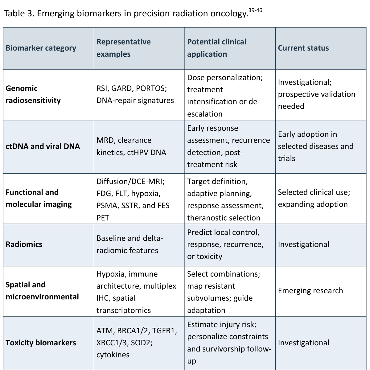
ctDNA is especially attractive when radiation aims to eradicate clinically occult disease. Clearance during or after radiation may provide early evidence of response; persistent ctDNA, tumor-informed molecular residual disease, or circulating viral DNA may identify recurrence risk before imaging. But the same caution applied elsewhere in this review applies here: a prognostic signal is not automatically an instruction to intensify or de-escalate radiation. Treatment changes should occur within validated, disease-specific pathways or prospective trials.16-19,43,44
Precision oncology is also moving beyond tumor-cell genomics to the geography of the tumor microenvironment. Multiplex immunohistochemistry or immunofluorescence, spatial transcriptomics, and digital pathology can map hypoxia, immune infiltration, T-cell exclusion, cancer-associated fibroblasts, angiogenesis, and stromal architecture within a treatment field. That spatial context matters because radiation, immunotherapy, and antibody-drug conjugates interact with both tumor cells and their surrounding ecosystem. The challenge is to convert a fascinating map into a reproducible result linked to a defined decision.45,46
Theranostics collapses diagnosis and treatment into the same molecular target. Imaging first demonstrates that the target is present and sufficiently distributed; a paired radioligand then delivers therapy to target-expressing cells. Somatostatin receptor imaging with lutetium-177 dotatate in neuroendocrine tumors and PSMA PET with lutetium-177 PSMA-617 in prostate cancer show how a biomarker can serve simultaneously as a selection tool, a map of disease, and a therapeutic gateway. FES PET and other molecular imaging approaches extend the same principle by revealing biologically meaningful target expression across the whole patient rather than in one biopsy.47,48
Precision radiation oncology is therefore moving from a fixed plan toward a living plan—one that integrates genomics, functional imaging, ctDNA, spatial biology, and patient-specific toxicity risk to select, adapt, intensify, or de-escalate treatment. Relatively few biomarkers currently direct routine radiation decisions, but the field is advancing quickly and belongs at the center, not the margins, of precision oncology.43,44
Antibody-drug conjugates (ADCs) challenge the conventional idea that target expression alone determines benefit. An ADC must bind its antigen, be internalized or release payload in the relevant microenvironment, deliver an active cytotoxin, and overcome cellular resistance. Activity can therefore depend on antigen abundance and spatial distribution, internalization kinetics, antibody affinity, linker stability, drug-to-antibody ratio, payload class and potency, membrane permeability, bystander effect, lysosomal processing, efflux pumps, DNA-repair capacity, and prior exposure to related payloads.20-23
HER2 provides the clearest example of how ADCs are rewriting the biomarker rulebook. Traditional testing was designed to identify tumors with amplification or protein overexpression likely to benefit from earlier HER2-directed therapies. DESTINY-Breast04 showed that trastuzumab deruxtecan improved outcomes in metastatic breast cancers classified as HER2-low. DESTINY-Breast06 then extended benefit to hormone receptor-positive HER2-low disease before chemotherapy and showed activity in a prespecified HER2-ultralow group.20,21 The lesson is not that every trace of HER2 is equivalent. It is that a potent ADC with a membrane-permeable payload and bystander effect can exploit expression below the threshold required for earlier HER2 treatments.
The DAISY study further demonstrated a gradient of activity across HER2 expression and provided mechanistic evidence that resistance may involve decreased HER2, impaired internalization, or payload-related biology.23 At the same time, reliable discrimination among IHC 0, ultralow, and 1+ disease is challenging, particularly in archival or heterogeneous specimens. Assay precision at the low end of expression becomes clinically consequential when a treatment indication depends on those categories.24
Other ADC targets—including folate receptor alpha, Nectin-4, TROP2, HER3, and B7-H3—show different relationships between expression and activity. For some approved ADCs, a companion diagnostic is required; for others, treatment is not restricted by a target-expression test. This apparent paradox reflects the totality of ADC biology and the design of the registrational trials. Future biomarkers may need to be quantitative, spatial, and functional, combining antigen density with heterogeneity, internalization, payload sensitivity, and resistance mechanisms rather than relying on a single positive/negative stain.22
Most oncology data are collected during a visit. Cancer and treatment toxicity, of course, do not wait for the next appointment. Digital biomarkers can help close that gap through objective measures from wearable accelerometers, connected sensors, smartphones, and other devices.25-27 Step count, activity intensity, gait speed, sleep timing, resting heart rate, respiratory rate, oxygen saturation, temperature, weight, and mobility patterns can complement performance status by showing how function changes in daily life—not only how a patient appears during a short clinic encounter.
Terminology matters. A patient-reported symptom entered into an app is an electronic patient-reported outcome (ePRO), not necessarily a digital biomarker, because it reflects the patient's direct report rather than an objective sensor-derived measure.27 Both can be clinically valuable. Randomized studies of electronic symptom monitoring have shown improvements in symptom control, quality of life, physical function, and selected measures of health care use; a single-center trial also reported longer survival, while the larger community-based PRO-TECT trial did not show an overall survival advantage despite benefits in patient-reported outcomes and fewer emergency visits.28-32 These mixed findings are instructive: the value lies not in digitizing a questionnaire alone, but in the complete intervention—patient participation, reliable alerts, accountable clinical response, and integration into care.
Wearable-derived signals could identify declining activity before a scheduled visit, quantify recovery after surgery, or help distinguish radiographic response from worsening functional status. A falling step count, rising resting heart rate, or disrupted sleep pattern may signal toxicity, infection, deconditioning, symptom burden, or disease progression. None is specific to cancer. Their usefulness depends on baseline calibration, longitudinal change, missing-data handling, and a response algorithm that avoids both missed deterioration and excessive false alarms.
Digital inclusion is part of validity, not a separate social consideration. Device ownership, broadband access, language, health literacy, manual dexterity, cognition, skin tone effects on optical sensors, and willingness to be monitored can influence who contributes usable data. If an algorithm is developed mainly in younger, affluent, digitally experienced populations, performance may degrade in the older and more medically complex patients who could benefit most. Validation should therefore include diverse populations, multiple devices, real-world adherence, and transparent reporting of missingness.
Oncology has no shortage of data. The bottleneck is turning those data into a pattern a clinical team can recognize and use. Artificial intelligence (AI) can extract relationships that are too high-dimensional for unaided interpretation. In radiomics, algorithms quantify tumor shape, texture, intensity, and spatial heterogeneity from CT, MRI, or PET images.33 In digital pathology, models can classify tissue, count cells, map tumor-infiltrating lymphocytes, assess spatial relationships, and infer selected molecular features from whole-slide images. Proof-of-concept studies have predicted common driver mutations from lung cancer histology, inferred multi-omic features and prognosis in colorectal cancer, and identified the likely origin of cancers of unknown primary.34-36
Radiomics can function as a biomarker in its own right, not merely as an input to an AI model. Baseline features may help estimate local control, pathologic response, nodal disease, immunotherapy response, or normal-tissue toxicity; delta-radiomics asks whether changes during treatment carry more information than the starting image. As with molecular assays, the relevant question is not how many features a model can extract, but whether a locked, validated result changes a decision.33,43-45
These approaches can create new biomarkers or improve the reproducibility of existing ones. An image-based model might flag tumors likely to be MSI-H for confirmatory testing, quantify HER2 heterogeneity across a slide, or characterize an immune-excluded phenotype. Multimodal models could combine pathology, radiology, genomics, ctDNA kinetics, laboratory values, treatment history, ePROs, and wearable data to estimate response or toxicity risk. The scientific appeal is obvious: cancer is a systems disease, and no single data stream captures the entire tumor-host interaction.2,33
The barriers are equally important. Retrospective models can learn site-specific staining, scanner, acquisition, or documentation patterns rather than biology. Performance often falls when a model is tested at an external center or in a later time period. Data leakage, inconsistent endpoints, class imbalance, and selective reporting can inflate apparent accuracy. A model that outputs a risk score without explaining intended use, uncertainty, and the action that should follow may be unsuitable for high-stakes care even if its area under the receiver operating characteristic curve is impressive.
Clinical readiness therefore requires a locked model, representative training data, external and temporal validation, assessment across demographic and technical subgroups, prospective workflow testing, monitoring for drift, and human oversight. The benchmark should not be whether AI can match a retrospective label. It should be whether the AI-enabled biomarker improves the accuracy, timeliness, consistency, or equity of a real clinical decision.
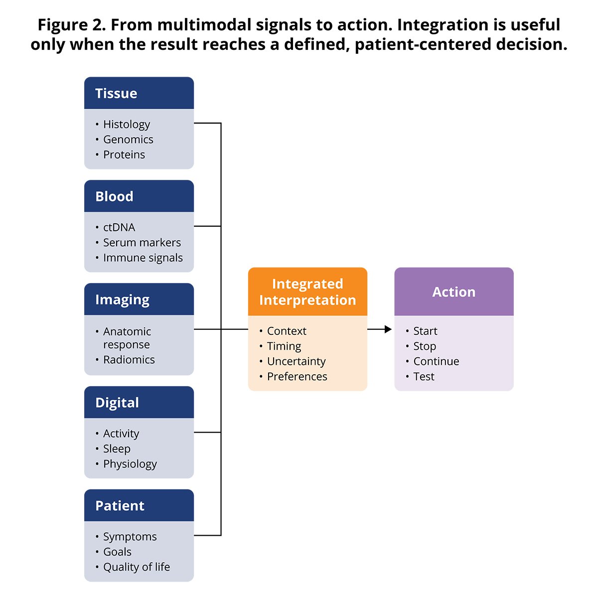
The emerging model follows the full cancer-care timeline. At diagnosis, tissue morphology, immunohistochemistry, and genomic profiling establish tumor identity and actionable biology. During treatment, imaging, serum markers, ctDNA kinetics, symptoms, and functional measures assess response and tolerability. After curative-intent therapy, molecular residual disease may refine recurrence risk. At progression, repeat tissue or plasma testing can reveal resistance mechanisms and new targets. During survivorship, symptoms, organ function, and digital measures may identify late effects or functional decline.
Integration does not mean placing every available data point on the clinician's screen. It means selecting complementary signals, defining how discordance will be resolved, and presenting information at the moment a decision is made. A practical system might combine a molecular trend, radiographic trajectory, toxicity signal, and functional trajectory into a concise decision view. The oncologist would still interpret the evidence in light of treatment goals, comorbidities, patient preferences, and uncertainty.
Discordant results will be common. A patient may have falling ctDNA but stable imaging, radiographic improvement but declining activity, or a targetable alteration in plasma that is absent from an older tissue specimen. The appropriate response depends on assay performance, timing, tumor shedding, lesion-specific biology, and the consequences of acting incorrectly. Multidisciplinary molecular tumor boards and clear institutional pathways can help adjudicate complex cases, but the field also needs prospective studies designed around discordance rather than treating it as noise.
The required evidence should match the stakes. An exploratory biomarker used to stratify a trial can tolerate more uncertainty than a test used to omit curative therapy. Evidence should progress from analytical validation to retrospective clinical association, external validation, and prospective demonstration of utility. For treatment-selection biomarkers, the most persuasive evidence shows a differential treatment effect according to biomarker status. For monitoring biomarkers, trials should show that acting on the signal improves outcomes or safely reduces unnecessary treatment, testing, or visits.
Assays require reference standards, proficiency testing, transparent thresholds, and consistent reporting. Longitudinal care also depends on interoperability: pathology, genomics, imaging, laboratory, ePRO, and wearable data must be available in compatible formats and attached to the correct clinical context. Without structured workflows, more testing can create more fragmentation rather than more precision.
Precision oncology can widen disparities if comprehensive testing, high-quality tissue, subspecialty interpretation, targeted therapies, and digital tools are concentrated in well-resourced centers. Equity should be measured across the entire pathway: who is offered testing, whose sample is adequate, who receives results before a decision, who can afford the recommended therapy, and whose data are represented in validation cohorts. A technically elegant biomarker that is unavailable or unactionable for much of the intended population has limited real-world utility.
Serial biomarkers can generate false alarms, incidental findings, anxiety, and downstream procedures. Digital monitoring can overwhelm teams if alerts are not prioritized and assigned. Every monitoring program should specify the frequency of measurement, clinically meaningful change, confirmatory step, responsible team member, response time, and escalation pathway. The operational design is part of the intervention and should be evaluated alongside the assay.
Patients should understand what a biomarker can and cannot answer, why repeated testing is being performed, how uncertain or discordant findings will be handled, and whether results could have hereditary implications. In digital programs, consent should address what data are collected, who can see them, how long they are retained, and what level of real-time monitoring the clinical team can realistically provide. Patient-centered design is essential to adherence, trust, and clinical relevance.
Conclusion: better signals, better decisionsBiomarkers have already transformed oncology by helping identify the right treatment for the right patient. Their next contribution will be broader: detecting response earlier, identifying molecular residual disease and recurrence, revealing emerging resistance, personalizing treatment intensity, anticipating selected toxicities, and showing how patients function between visits.
The next era will not be defined by one dominant biomarker. It will be defined by disciplined integration of molecular, pathologic, imaging, spatial, clinical, physiologic, and patient-reported signals into decisions that are timely, interpretable, equitable, and demonstrably useful. The next generation of biomarkers will not simply identify who should receive a therapy. They will increasingly determine when treatment should begin, how much is needed, how long it should continue, and when it can safely be de-escalated. Precision oncology is moving from treatment selection toward continuous treatment adaptation across the course of disease.
The field's success should be judged not by how much data it can generate, but by whether those data help patients live longer or better—with less unnecessary treatment and greater confidence in the decisions made along the way.
1. FDA-NIH Biomarker Working Group. BEST (Biomarkers, EndpointS, and other Tools) Resource [Internet]. Silver Spring (MD): U.S. Food and Drug Administration; Bethesda (MD): National Institutes of Health; 2016-. Updated January 7, 2025. Accessed August 4, 2026. https://www.ncbi.nlm.nih.gov/books/NBK326791/
2. Passaro A, Al Bakir M, Hamilton EG, et al. Cancer biomarkers: emerging trends and clinical implications for personalized treatment. Cell. 2024;187(7):1617-1635. doi:10.1016/j.cell.2024.02.041
3. U.S. Food and Drug Administration. List of Cleared or Approved Companion Diagnostic Devices (In Vitro and Imaging Tools). Accessed August 4, 2026. https://www.fda.gov/medical-devices/in-vitro-diagnostics/list-cleared-or-approved-companion-diagnostic-devices-in-vitro-and-imaging-tools
4. National Cancer Institute. Biomarker Testing for Cancer Treatment. Updated December 14, 2021. Accessed August 4, 2026. https://www.cancer.gov/about-cancer/treatment/types/biomarker-testing-cancer-treatment
5. Kim J, Kim HS, Chae YK. Tissue-agnostic biomarkers in solid tumors: current approvals and emerging candidates. Cancer Metastasis Rev. 2025;44:58. doi:10.1007/s10555-025-10274-2
6. Soria JC, Ohe Y, Vansteenkiste J, et al. Osimertinib in untreated EGFR-mutated advanced non-small-cell lung cancer. N Engl J Med. 2018;378(2):113-125. doi:10.1056/NEJMoa1713137
7. Peters S, Camidge DR, Shaw AT, et al. Alectinib versus crizotinib in untreated ALK-positive non-small-cell lung cancer. N Engl J Med. 2017;377(9):829-838. doi:10.1056/NEJMoa1704795
8. Drilon A, Laetsch TW, Kummar S, et al. Efficacy of larotrectinib in TRK fusion-positive cancers in adults and children. N Engl J Med. 2018;378(8):731-739. doi:10.1056/NEJMoa1714448
9. Andre T, Shiu KK, Kim TW, et al. Pembrolizumab in microsatellite-instability-high advanced colorectal cancer. N Engl J Med. 2020;383(23):2207-2218. doi:10.1056/NEJMoa2017699
10. Le DT, Durham JN, Smith KN, et al. Mismatch repair deficiency predicts response of solid tumors to PD-1 blockade. Science. 2017;357(6349):409-413. doi:10.1126/science.aan6733
11. Rimm DL, Han G, Taube JM, et al. A prospective, multi-institutional, pathologist-based assessment of 4 immunohistochemistry assays for PD-L1 expression in non-small cell lung cancer. JAMA Oncol. 2017;3(8):1051-1058. doi:10.1001/jamaoncol.2017.0013
12. Marabelle A, Fakih M, Lopez J, et al. Association of tumour mutational burden with outcomes in patients with advanced solid tumours treated with pembrolizumab: prospective biomarker analysis of the multicohort, open-label, phase 2 KEYNOTE-158 study. Lancet Oncol. 2020;21(10):1353-1365. doi:10.1016/S1470-2045(20)30445-9
13. Eisenhauer EA, Therasse P, Bogaerts J, et al. New response evaluation criteria in solid tumours: revised RECIST guideline (version 1.1). Eur J Cancer. 2009;45(2):228-247. doi:10.1016/j.ejca.2008.10.026
14. Seymour L, Bogaerts J, Perrone A, et al. iRECIST: guidelines for response criteria for use in trials testing immunotherapeutics. Lancet Oncol. 2017;18(3):e143-e152. doi:10.1016/S1470-2045(17)30074-8
15. National Cancer Institute. Tumor Marker Tests in Common Use. Updated December 7, 2023. Accessed August 4, 2026. https://www.cancer.gov/about-cancer/diagnosis-staging/diagnosis/tumor-markers-list
16. Tie J, Wang Y, Lo SN, et al. Circulating tumor DNA analysis guiding adjuvant therapy in stage II colon cancer: 5-year outcomes of the randomized DYNAMIC trial. Nat Med. 2025;31(5):1509-1518. doi:10.1038/s41591-025-03579-w
17. Nakamura Y, Watanabe J, Akazawa N, et al. ctDNA-based molecular residual disease and survival in resectable colorectal cancer. Nat Med. 2024;30(11):3272-3283. doi:10.1038/s41591-024-03254-6
18. Powles T, Assaf ZJ, Davarpanah N, et al. ctDNA guiding adjuvant immunotherapy in urothelial carcinoma. Nature. 2021;595(7867):432-437. doi:10.1038/s41586-021-03642-9
19. Emiloju OE, Storandt M, Zemla T, et al. Tumor-informed circulating tumor DNA for minimal residual disease detection in the management of colorectal cancer. JCO Precis Oncol. 2024;8:e2300127. doi:10.1200/PO.23.00127
20. Modi S, Jacot W, Yamashita T, et al. Trastuzumab deruxtecan in previously treated HER2-low advanced breast cancer. N Engl J Med. 2022;387(1):9-20. doi:10.1056/NEJMoa2203690
21. Bardia A, Hu X, Dent R, et al. Trastuzumab deruxtecan after endocrine therapy in metastatic breast cancer. N Engl J Med. 2024;391(22):2110-2122. doi:10.1056/NEJMoa2407086
22. Tarantino P, Carmagnani Pestana R, Corti C, et al. Antibody-drug conjugates: smart chemotherapy delivery across tumor histologies. CA Cancer J Clin. 2022;72(2):165-182. doi:10.3322/caac.21705
23. Mosele F, Deluche E, Lusque A, et al. Trastuzumab deruxtecan in metastatic breast cancer with variable HER2 expression: the phase 2 DAISY trial. Nat Med. 2023;29(8):2110-2120. doi:10.1038/s41591-023-02478-2
24. Wolff AC, Somerfield MR, Dowsett M, et al. Human epidermal growth factor receptor 2 testing in breast cancer: ASCO-College of American Pathologists guideline update. J Clin Oncol. 2023;41(22):3867-3872. doi:10.1200/JCO.22.02864
25. Macias Alonso AK, Hirt J, Woelfle T, Janiaud P, Hemkens LG. Definitions of digital biomarkers: a systematic mapping of the biomedical literature. BMJ Health Care Inform. 2024;31(1):e100914. doi:10.1136/bmjhci-2023-100914
26. U.S. Food and Drug Administration. Digital Health Technologies for Remote Data Acquisition in Clinical Investigations: Guidance for Industry, Investigators, and Other Stakeholders. December 2023. Accessed August 4, 2026. https://www.fda.gov/regulatory-information/search-fda-guidance-documents/digital-health-technologies-remote-data-acquisition-clinical-investigations
27. Izmailova ES, Demanuele C, McCarthy M. Digital health technology-derived measures: biomarkers or clinical outcome assessments? Clin Transl Sci. 2023;16(7):1113-1120. doi:10.1111/cts.13529
28. Basch E, Deal AM, Kris MG, et al. Symptom monitoring with patient-reported outcomes during routine cancer treatment: a randomized controlled trial. J Clin Oncol. 2016;34(6):557-565. doi:10.1200/JCO.2015.63.0830
29. Basch E, Deal AM, Dueck AC, et al. Overall survival results of a trial assessing patient-reported outcomes for symptom monitoring during routine cancer treatment. JAMA. 2017;318(2):197-198. doi:10.1001/jama.2017.7156
30. Denis F, Lethrosne C, Pourel N, et al. Randomized trial comparing a web-mediated follow-up with routine surveillance in lung cancer patients. J Natl Cancer Inst. 2017;109(9):djx029. doi:10.1093/jnci/djx029
31. Basch E, Schrag D, Henson S, et al. Effect of electronic symptom monitoring on patient-reported outcomes among patients with metastatic cancer: a randomized clinical trial. JAMA. 2022;327(24):2413-2422. doi:10.1001/jama.2022.9265
32. Basch E, Schrag D, Jansen J, et al. Symptom monitoring with electronic patient-reported outcomes during cancer treatment: final results of the PRO-TECT cluster-randomized trial. Nat Med. 2025;31(4):1225-1232. doi:10.1038/s41591-025-03507-y
33. Lambin P, Leijenaar RTH, Deist TM, et al. Radiomics: the bridge between medical imaging and personalized medicine. Nat Rev Clin Oncol. 2017;14(12):749-762. doi:10.1038/nrclinonc.2017.141
34. Coudray N, Ocampo PS, Sakellaropoulos T, et al. Classification and mutation prediction from non-small cell lung cancer histopathology images using deep learning. Nat Med. 2018;24(10):1559-1567. doi:10.1038/s41591-018-0177-5
35. Lu MY, Chen TY, Williamson DFK, et al. AI-based pathology predicts origins for cancers of unknown primary. Nature. 2021;594(7861):106-110. doi:10.1038/s41586-021-03512-4
36. Tsai PC, Lee TH, Kuo KC, et al. Histopathology images predict multi-omics aberrations and prognoses in colorectal cancer patients. Nat Commun. 2023;14(1):2102. doi:10.1038/s41467-023-37179-4
37. Amstutz U, Henricks LM, Offer SM, et al. Clinical Pharmacogenetics Implementation Consortium guideline for dihydropyrimidine dehydrogenase genotype and fluoropyrimidine dosing: 2017 update. Clin Pharmacol Ther. 2018;103(2):210-216. doi:10.1002/cpt.911
38. U.S. Food and Drug Administration. Table of Pharmacogenomic Biomarkers in Drug Labeling. Updated March 3, 2026. Accessed August 4, 2026. https://www.fda.gov/drugs/science-and-research-drugs/table-pharmacogenomic-biomarkers-drug-labeling
39. Torres-Roca JF. A molecular assay of tumor radiosensitivity: a roadmap towards biology-based personalized radiation therapy. Per Med. 2012;9(5):547-557. doi:10.2217/pme.12.55
40. Scott JG, Berglund A, Schell MJ, et al. A genome-based model for adjusting radiotherapy dose (GARD): a retrospective, cohort-based study. Lancet Oncol. 2017;18(2):202-211. doi:10.1016/S1470-2045(16)30648-9
41. Eschrich S, Zhang H, Zhao H, et al. Systems biology modeling of the radiation sensitivity network: a biomarker discovery platform. Int J Radiat Oncol Biol Phys. 2009;75(2):497-505. doi:10.1016/j.ijrobp.2009.05.056
42. West CM, Barnett GC. Genetics and genomics of radiotherapy toxicity: towards prediction. Genome Med. 2011;3(8):52. doi:10.1186/gm268
43. Baumann M, Krause M, Overgaard J, et al. Radiation oncology in the era of precision medicine. Nat Rev Cancer. 2016;16(4):234-249. doi:10.1038/nrc.2016.18
44. Quon H, McNutt T, Lee J, et al. Needs and challenges for radiation oncology in the era of precision medicine. Int J Radiat Oncol Biol Phys. 2019;103(4):809-817. doi:10.1016/j.ijrobp.2018.11.017
45. Lewis SM, Asselin-Labat ML, Nguyen Q, et al. Spatial omics and multiplexed imaging to explore cancer biology. Nat Methods. 2021;18(9):997-1012. doi:10.1038/s41592-021-01203-6
46. Binnewies M, Roberts EW, Kersten K, et al. Understanding the tumor immune microenvironment (TIME) for effective therapy. Nat Med. 2018;24(5):541-550. doi:10.1038/s41591-018-0014-x
47. Strosberg J, El-Haddad G, Wolin E, et al. Phase 3 trial of 177Lu-Dotatate for midgut neuroendocrine tumors. N Engl J Med. 2017;376(2):125-135. doi:10.1056/NEJMoa1607427
48. Sartor O, de Bono J, Chi KN, et al. Lutetium-177-PSMA-617 for metastatic castration-resistant prostate cancer. N Engl J Med. 2021;385(12):1091-1103. doi:10.1
Rebecca Previs
 Rebecca Previs
 Ali Raza Khaki
 Avi Cherla, MSc; Huseyin Naci, MHS, PhD; Aaron S. Kesselheim, MD, JD, MPH; Bishal Gyawali, MD, PhD; Elias Mossialos, MD, PhD
 Michael Pietrack
Jamie Riley
Michael Pietrack
Jamie Riley